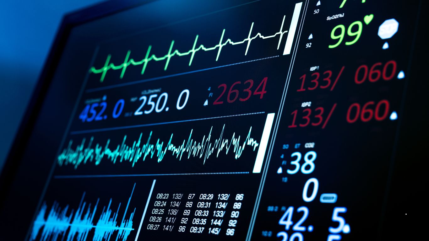
Understanding ECG readings—both normal and abnormal—provides crucial insights into cardiac health.
An electrocardiogram (ECG) traces the electrical activity of the heart, revealing its rhythm and detecting any irregularities. Interpreting these readings accurately can mean early detection of potential issues or reassurance of a healthy heart. From the distinctive peaks and valleys of a normal sinus rhythm to the nuanced deviations indicative of arrhythmias or ischemia, each waveform tells a story.
Whether you’re a healthcare professional striving for precision or a curious individual eager to delve deeper, explore the world of ECG readings and uncover the secrets of cardiac health. Dive in!
Basics Of Ecg Interpretation
Discover the basics of ECG interpretation with a focus on reading normal and abnormal electrocardiograms. Gain insights into understanding the intricacies of ECG patterns and how to identify potential issues for effective diagnosis.
Understanding ECG And Its Importance In Medicine
The electrocardiogram (ECG) is a crucial diagnostic tool used in the field of medicine to assess the electrical activity of the heart. It records the rhythmic electrical impulses that travel through the heart’s chambers, providing valuable insights into cardiac health and functioning.
Components Of A Normal Ecg Reading
A normal ECG reading comprises several key components that reflect the various stages of a heartbeat. Understanding these components is essential for accurate interpretation.
P waves:
- Atrial depolarization, represented by the P wave, signifies the contraction of the atria to pump blood into the ventricles.
PR interval:
- The PR interval represents the passage of electrical impulses from the atria to the ventricles, allowing for proper coordination of contractions.
QRS complex:
- The QRS complex signifies ventricular depolarization, the electrical activity associated with the contraction of the ventricles to propel blood into the arteries.
ST segment:
- The ST segment reflects the period between ventricular depolarization and repolarization, representing the initial phase of ventricular relaxation.
T waves:
- T waves represent ventricular repolarization, indicating the recovery and preparation of the heart for the next cycle of contraction.
QT interval:
- The QT interval represents the time duration of ventricular depolarization and repolarization, providing insights into potential abnormalities in cardiac repolarization.
Standard ECG Graph Explained
Understanding the standard ECG graph is crucial for accurate interpretation of ECG readings. The graph consists of multiple leads that capture electrical signals from specific positions on the body. These leads are classified into two main types:
- Limb Leads
- I, II, III: These leads record electrical activity between specific combinations of limbs, providing valuable information about the electrical axis of the heart and its orientation within the chest.
- aVR, aVL, aVF: These augmented leads record voltages from the heart using a combination of positive and negative electrode placements.
- Precordial (Chest) Leads
- V1-V6: These leads are placed on specific points of the chest wall to capture electrical signals corresponding to the heart’s activity from various angles.
By evaluating the electrical patterns captured by these leads, healthcare professionals can identify deviations from normal and recognize potential abnormalities or irregularities in the heart’s electrical activity. This information assists in diagnosing conditions such as arrhythmias, ischemia, myocardial infarctions, and other cardiac disorders.
Understanding the basics of ECG interpretation, including the components of a normal ECG reading and the standard ECG graph, is essential in accurately assessing cardiac health and function. By familiarizing yourself with the intricacies of ECG analysis, you can improve your understanding of cardiac physiology and contribute to more effective patient care.
Abnormal ECG: Spotting The Signs
Spotting the signs of abnormal ECG readings can help identify potential heart conditions. Learn about normal and abnormal ECG readings in this informative article.
Common Abnormalities Seen In ECG Readings
Interpreting ECG readings requires a keen eye to detect abnormalities. Here are some of the most commonly observed abnormalities that healthcare professionals might encounter:
1. Arrhythmias: Arrhythmias refer to irregular heartbeats that deviate from the normal sinus rhythm. They can manifest as tachycardia (rapid heart rate) or bradycardia (slow heart rate).
2. Myocardial Infarction (MI): A myocardial infarction, commonly known as a heart attack, can be detected through ECG readings. The presence of ST-segment elevation or depression signifies damage to the heart muscle.
3. Atrial Fibrillation: Atrial fibrillation is characterized by abnormal electrical signals originating from the atria, resulting in an irregular and often rapid heartbeat. This condition is typically identified by the absence of P waves and an irregularly irregular rhythm.
4. Conduction Blocks: Conduction blocks occur when the electrical signals between the chambers of the heart are delayed or blocked. These blocks can be categorized as first-degree, second-degree, or third-degree, depending on the severity of the interruption.
Significance Of Abnormal Waveforms And Intervals
Abnormal waveforms and intervals in an ECG reading provide valuable insights into the heart’s functioning and potential cardiovascular conditions. Let’s explore the significance of these abnormalities:
1. ST-segment elevation or depression: ST-segment deviations beyond the normal range may indicate a myocardial infarction or ischemia, indicating reduced blood flow to the heart muscle.
2. QT interval prolongation: A prolonged QT interval on the ECG suggests a delay in ventricular repolarization, which can increase the risk of serious arrhythmias and sudden cardiac arrest.
3. Prolonged PR interval: A prolonged PR interval could indicate an atrioventricular (AV) block, which disrupts the transmission of electrical signals between the atria and ventricles.
Correlating Symptoms With ECG Changes
Correlating symptoms with ECG changes is crucial to ensure accurate diagnosis and appropriate treatment. Symptoms that may correlate with abnormal ECG findings include: – Chest pain or discomfort – Shortness of breath – Dizziness or lightheadedness – Palpitations or irregular heartbeats – Fatigue or weakness – Fainting or syncope By analyzing the ECG in conjunction with the patient’s symptoms, healthcare professionals can better understand the underlying cardiac condition and provide timely interventions.
ECGs In Diagnosing Heart Conditions
Electrocardiograms (ECGs) play a crucial role in diagnosing heart conditions and are often the initial step taken by healthcare professionals when evaluating a patient’s cardiovascular health.
Role Of An ECG in Identifying Heart Diseases
The role of an ECG in identifying heart diseases cannot be overstated. It serves as a valuable tool for healthcare professionals in assessing the overall heart health of patients. By analyzing the electrical signals produced by the heart, an ECG can detect a wide range of heart conditions, including:
- Arrhythmias – irregular heart rhythms that may be too fast, too slow, or irregular
- Heart attacks – disruptions in blood flow to the heart muscle
- Coronary artery disease – blockages or narrowing of the blood vessels that supply the heart
- Heart valve problems – issues with the valves that regulate blood flow within the heart
- Cardiomyopathy – disease affecting the heart muscles
Distinguishing Between Urgent And Non-urgent Abnormalities
ECG readings can further aid in differentiating between urgent and non-urgent abnormalities. Not all abnormal readings on an ECG indicate an immediate threat to a patient’s health. Some abnormalities may be benign or require further observation, while others demand immediate medical attention.
Examples of urgent abnormalities that warrant immediate attention include:
- ST-segment elevation – a significant indicator of a heart attack
- Ventricular fibrillation – a life-threatening arrhythmia
- Complete heart block – a blockage in the electrical signals between the upper and lower chambers of the heart
On the other hand, non-urgent abnormalities, such as minor arrhythmias or isolated premature ventricular contractions (PVCs), may not require immediate intervention but could necessitate regular monitoring or further evaluation.
Case Studies: Ecg Readings In Different Cardiac Scenarios
Let’s delve into some case studies that highlight the utility of ECG readings in different cardiac scenarios:
| Case | ECG Finding | Diagnosis |
|---|---|---|
| Case 1 | ST-segment elevation | Acute myocardial infarction (heart attack) |
| Case 2 | Prolonged QT interval | Long QT syndrome |
| Case 3 | Absent P waves | Atrial fibrillation |
These case studies demonstrate how ECG readings provide critical diagnostic information, enabling healthcare professionals to determine the appropriate course of action for each patient.
From ECG To Action: Clinical Response
In this section, I will delve into each of these aspects in detail, providing you with a comprehensive understanding of how to translate ECG findings into actionable clinical responses.
Interpreting ECG findings: Normal variants vs. pathologyThe interpretation of ECG findings can be challenging, as it requires distinguishing between normal variants and pathological abnormalities. Normal variants are variations in the ECG waveform that are typically not associated with any underlying cardiac disease or dysfunction.
Some common normal variants that can be observed on an ECG include:
| Normal Variant | Description |
|---|---|
| Sinus arrhythmia | An irregular beat-to-beat interval variation, is usually associated with respiration. |
| Early repolarization | ST-segment elevation at the J point, is often seen in young individuals. |
| High QRS voltage | An increase in the amplitude of the QRS complex, which can be caused by certain conditions but is also seen in athletes. |
Pathological findings, on the other hand, indicate underlying cardiac abnormalities or diseases that require further investigation and management.
Examples of pathological findings include:
- ST-segment elevation myocardial infarction (STEMI)
- Bundle branch block (BBB)
- Atrial fibrillation (AF)
Guidelines For Immediate Management Based On ECG Results
Once an ECG reading has been obtained, it is essential to determine the appropriate management strategy promptly. The management plan will depend on the specific findings observed in the ECG, whether they are normal variants or pathological abnormalities.
For normal variants, no immediate intervention is typically necessary. However, it is crucial to document these findings in the patient’s medical record for future reference.
If the ECG shows pathological findings indicative of a potentially life-threatening condition, immediate action must be taken. This may involve:
- Notifying the appropriate medical personnel to evaluate the patient’s condition.
- Implementing emergency measures such as cardiac monitoring, administering medications, or performing cardioversion.
- Preparing the patient for further investigations or interventions, such as coronary angiography or cardiac surgery.
Follow-up Investigations After An Abnormal ECG Reading
The specific investigations required will depend on the suspected diagnosis based on the ECG findings. Some common follow-up investigations may include:
- Echocardiography: To assess cardiac structure and function.
- Cardiac stress test: To evaluate the heart’s response to exercise or stress.
- Coronary angiography: To visualize the coronary arteries and identify any blockages or narrowing.
- Electrophysiological studies: To evaluate the electrical conduction system of the heart and identify any abnormalities.
- Genetic testing: To identify any underlying genetic conditions or predispositions.
Beyond The Beats: ECG’s Broader Context
While many of us are familiar with ECG or electrocardiogram as a tool used for monitoring heart health, it’s important to recognize its broader context beyond just the beats. ECG readings can provide valuable insights into a person’s overall heart health, detecting both normal and abnormal rhythms.
Impact Of Lifestyle On ECG Readings Normal And Abnormal
Your lifestyle choices play a vital role in determining the accuracy of your ECG readings. Various factors such as smoking, excessive alcohol consumption, stress, and lack of physical activity can contribute to abnormal ECG patterns.
Additionally, certain medications and underlying medical conditions can also influence the interpretation of ECG results. By adopting a healthy lifestyle, which includes regular exercise, a balanced diet, stress management, and avoiding harmful habits, you can positively impact your ECG readings.
Future Of ECG Technology In Heart Health Monitoring
The constant advancements in technology have paved the way for exciting opportunities in ECG monitoring. With the emergence of wearable devices, individuals can now conveniently track their heart health in real time, providing a holistic view of their cardiovascular well-being.
These innovative devices, equipped with advanced algorithms and high-precision sensors, allow for continuous monitoring of heart rate variability, early detection of arrhythmias, and the ability to instantly share ECG data with healthcare professionals. The future holds immense potential for ECG technology to revolutionize heart health monitoring and improve early detection, ultimately leading to better outcomes for patients.
Encouraging Heart Health Awareness Through ECG Education
One of the key components of harnessing the full potential of ECG technology is education and awareness. By providing comprehensive information and resources about ECGs, individuals can better understand the significance of their readings and take proactive steps toward maintaining a healthy heart.
Promoting heart health awareness through ECG education can involve initiatives such as workshops, online courses, and the sharing of accurate and accessible information through various media channels. By empowering individuals with knowledge about their heart health, we can collectively strive towards a healthier society.
Conclusion
Understanding the difference between normal and abnormal ECG readings is crucial for diagnosing and treating cardiac conditions. By recognizing the patterns and anomalies in the ECG tracings, healthcare professionals can accurately interpret the data and make informed decisions about patient care.
It is important to prioritize regular ECG screenings to ensure early detection and proactive management of any potential heart health issues. Stay vigilant and proactive in monitoring your heart health for a better quality of life.
FAQs Of Ecg Reading Normal And Abnormal
What Is An ECG Reading?
An ECG reading, also known as an electrocardiogram, is a test that measures the electrical activity of the heart. It helps detect abnormalities in heart rhythm, such as irregularities, blockages, or damage to the heart muscle.
How Is A Normal Ecg Reading Interpreted?
A normal ECG reading shows a regular heart rhythm, consistent intervals, and no signs of abnormalities. The P wave represents atrial depolarization, the QRS complex represents ventricular depolarization, and the T wave represents ventricular repolarization. A normal ECG reading indicates a healthy heart.
What Are Some Common Abnormalities In An ECG Reading?
Common abnormalities in an ECG reading include abnormal heart rhythms, such as atrial fibrillation or ventricular tachycardia, ST-segment elevation or depression indicating an ischemic event, or Q-wave abnormalities indicating a previous heart attack.
How Can An ECG Reading Help Detect Heart Conditions?
An ECG reading can help detect various heart conditions by identifying irregular heart rhythms, signs of coronary artery disease, heart muscle damage, or problems with the heart’s electrical system. It is a valuable tool for diagnosing conditions such as heart attacks, arrhythmias, or ischemic events.

Nazmul Gazi is a dedicated final-year student at Cumilla Medical College with a passion for promoting health and wellness. Drawing from his medical studies, Nazmul writes insightful health tips and guides, helping readers make informed decisions about their well-being.

